
Figure 3. Inhibitory effect of i-lncRNA on cell cycle and apoptosis.
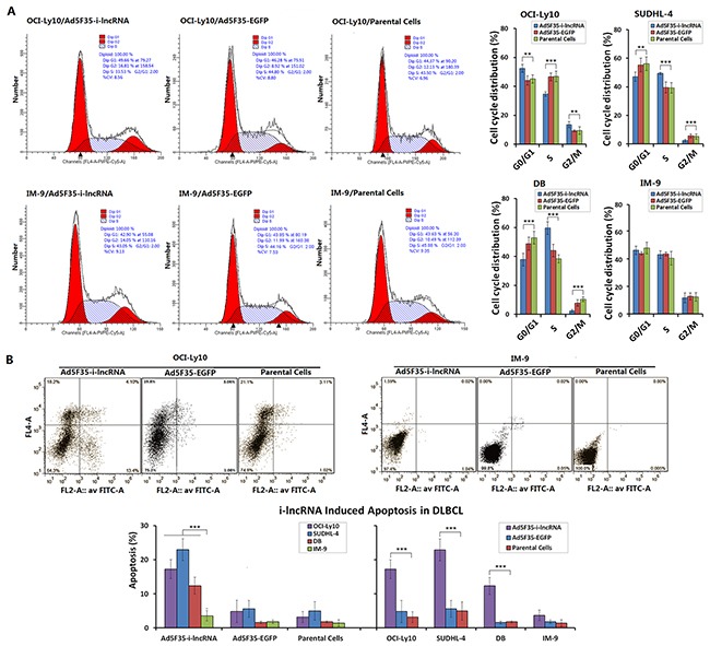
A. The four cell lines were cultured in 6-well plates at a density of 1 × 10⁵ cells/100 μL/well for 24 h, then infected with Ad5F35-i-lncRNA or Ad5F35-EGFP at an MOI of 100 pfu/cell. After continuously cultured for 48 h, cells were harvested and fixed in pre-chilled 75% ethanol, placed in a 4°C refrigerator overnight, washed in PBS twice, added the RNase-containing PI staining mixture and incubated in dark for 30 min. Cells were subjected to cell cycle analysis by flow cytometry; **P<0.01 and ***P<0.001. B. Cells were cultured and infected with the viruses as aforementioned, harvested 48 h later and stained with Annexin V/PI. Cell apoptosis was analyzed by flow cytometric analysis; ***P<0.001.