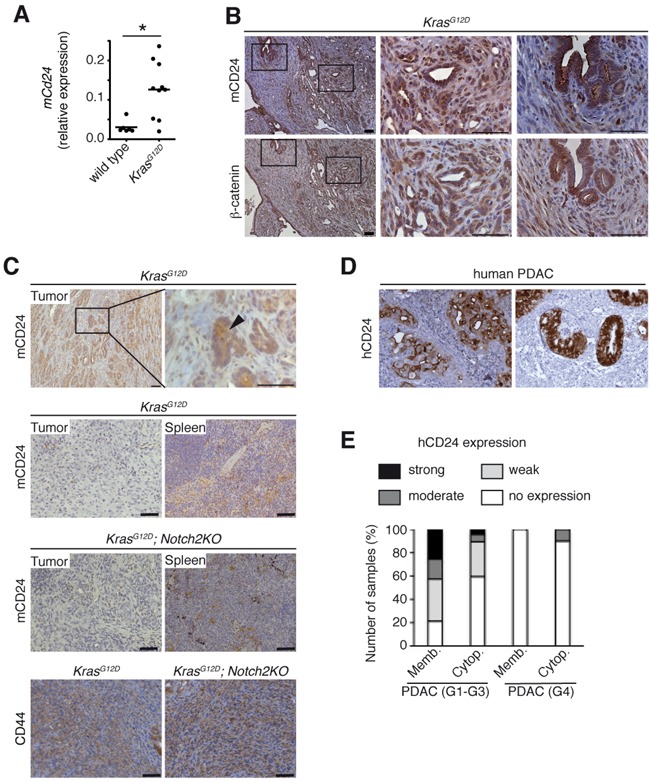
Figure 1. h/mCD24 is expressed in differentiated PDAC.

A. Cd24 mRNA expression increased in KrasG12D mice compared to wild type at the age of 6 months. B. Immunohistological staining for mCD24 and β-catenin in murine differentiated PDAC. C. Immunohistological staining of murine differentiated and undifferentiated PDAC of indicated genotypes. Spleens from the same mice were used as a positive control for staining. D. Immunohistological staining for hCD24 in human PDAC. Examples showing membranous, cytoplasmic or both staining patterns. E. A series of human ductal (G1-G3) and undifferentiated (G4) pancreatic cancers was stained for hCD24. The membrane-bound and cytoplasmic stainings were scored according to a three-tiered system (1 - <10%, 2 - 11-50%, 3 - >50% of the cells are positive). Scale bars = 50 μM.
