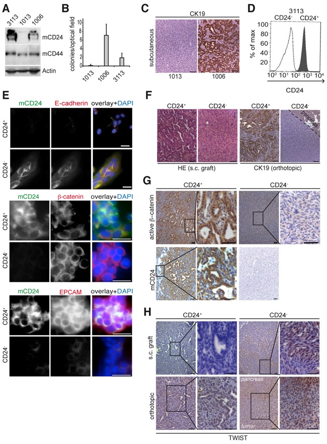
Figure 2. mCD24 expressing tumor cells lead to differentiated tumors.

Pancreatic cells were either mCD24 negative (#1006) or mCD24 positive (#1006 and #3113). A. Western blot analysis of primary mouse cell lines. B. Soft agar assay. Mean ± SD. C. immunohistological analysis of s.c. tumors generated from CD24− or CD24+ pancreatic cells. D, E. Pancreatic cells (#3113) were sorted into CD24− and CD24+ cell populations. D, purity of sorted populations was confirmed by FACS analysis using a mCD24-FITC antibody. E, immunofluorescence staining of sorted cells. F. Left panels: histology of tumors (N=13) generated by s.c. injections of CD24− and CD24+ pancreatic cells. Right panels: immunohistological analysis for CK19 expression of tumors generated by orthotopic transplantation of CD24− and CD24+ pancreatic cells. G, H. Immunohistological analysis of s.c. and orthotopic tumors, as described in F, showing strong cytoplasmic expression of active β-catenin in CD24+ tumors and nuclear TWIST expression in CD24- tumors. Scale bars = 50 μm.
