
Figure 1. Synergisitic effects of IFN-α and IFN-λ on in vivo BNL tumor growth.
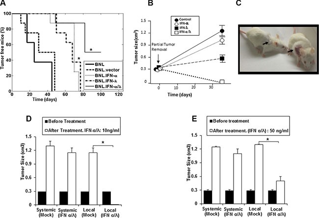
(A) Syngeneic BALB/c mice (n = 8) were injected s.c. in the flank with 106 BNL, BNL.vector, BNL.IFN-λ, BNL.IFN-α cells, or a 50:50 combination of both IFN-producing cells. Data in the Kaplan-Meier survival curves are shown as percentage of tumor free mice. BNL.IFN-α or BNL.IFN-λ versus BNL.IFN-α/λ cells (*) p < 0.05 (one-way ANOVA). (B) Mice (n = 5 per group) were inoculated with 106 parental BNL cells at the back. After tumor growth (1 cm3), partial tumor removal was performed (0.3 cm3 left). Mice were then treated with 10 ng of either IFN-α or IFN-λ alone, or a combination of IFN-α and IFN-λ (50% of each dose) at the tumor surgery site, starting two days post-surgery, three days a week for two weeks; and tumor growth was monitored. Data are presented as the mean tumor volume ± SE (n = 5). (C) Representative example showing a mouse, treated with IFN-α and IFN-λ combination (Left) and control (Right). Arrows indicate the tumor site and area of treatment with IFN-α and IFN-λ combination or control (Mock). (D and E) Mice (n = 5 per group) were inoculated with 106 parental BNL cells at the neck. When the tumor reached around 1 cm3, partial tumor removal was performed. Mice were then treated locally at the tumor inoculation site at the neck or systemically (i. p) with either 10 ng (d) or 50 ng (e) of a combination of IFN-α and IFN-λ (50% of each IFN dose), three days a week for two weeks and starting two days post-surgery. Experiments are repeated 3 times and data presented as the mean tumor volume ± SE. (*) p < 0.05. p value determined by Mann-Whitney U test.