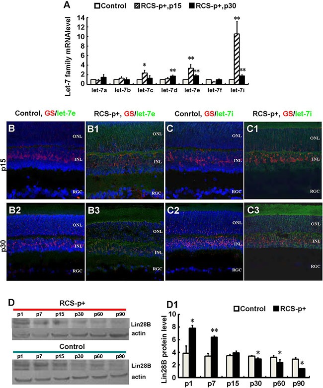
Figure 3. Upregulateion of let-7e and let-7i and downregulation of Lin28B in dystrophic rat retinas.

(A) Relative quantitative analysis showed that most members of the let-7 family, except let-7a and let-7f at p15, were upregulated at p15 and p30 in RCS-p+ rats' retina compared with controls. Among these members, let-7e and let-7i were upregulated most obviously. (B–B3 and C–C3) Immunofluorescence simultaneously stained against GS (red) and in situ hybridization with LNA probes against let-7e or let-7i (green). The expression of let-7e and let-7i co-localized with GS in somas and processes of Müller cells. The intensities of these two molecular signals in RCS-p+ rat retinas were stronger than that of controls at early p15 and p30. (D–D1) Western blotting analysis showed that the expression of Lin28B protein only increased before retinal degeneration at p1 and p7, then was reduced after retinal degeneration at p15 in RCS-p+ rat retinas when compared with control rat retinas. Representative results are shown. Data are presented as the mean ± standard error from three replicates. *P < 0.05, **P < 0.01, Student's t-test.
