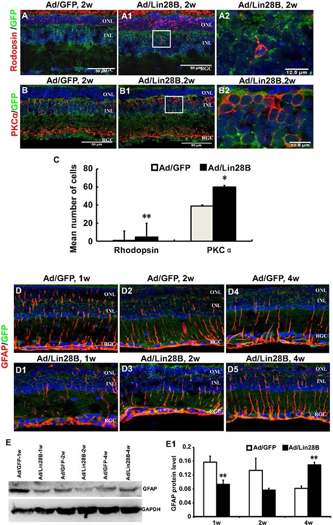
Figure 6. Ectopic Lin28B expression promotes neurogenesis and inhibits gliogenesis of de-differentiated Müller cells in vivo.

(A–B2) Immunofluorescence staining against retinal neural markers rhodopsin and PKCα at 2 week after subretinal space injection of Ad/Lin 28B or Ad/GFP (green) in RCS-p+ rats. A2 and B2 are enlargements of the square in of A1–B1. (C) Quantitative and statistical analysis showed that the positive cells of rhodopsin and PKCα in the Ad/Lin28B infected group were significantly increased when compared with the Ad/GFP infected group. (D–D5) Immunofluorescence staining against GFAP at 1 week, 2 weeks, and 4 weeks after subretinal space injection of Ad/Lin 28B or Ad/GFP (green) in RCS-p+ rats. The result showed that the level of GFAP expression gradually decreased in Ad/Lin 28B treated retinas as compared to the retinas treated with Ad/GFP. (E–E1) Western blot analysis supported the result of immunofluorescence staining. Representative results are shown. Data are presented as the mean ± standard error from three replicates. *P < 0.05, **P < 0.01, Student's t-test.
