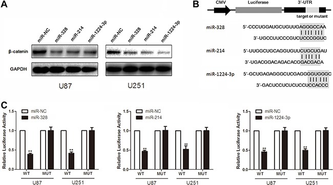
Figure 3. MiR-328, miR-214 and miR-1224-3p target β-catenin.

(A) Western blot analysis of lysates from cells transfected by miR-328, miR-214 or miR-1224-3p probed with β-catenin antibody. GAPDH was served as the loading control. (B) Schematic representation of the putative binding sites in β-catenin mRNAs 3′UTR for miR-328, miR-214 and miR-1224-3p. (C) pGL3-WT-β-catenin-3′UTR-Luc and pGL3-MUT-β-catenin-3′UTR-Luc reporters were transfected into glioma cells treated by miR-328, miR-214 or miR-1224-3p. Luciferase activity was determined 48 h after transfection. The ratio of normalized sensor to control luciferase activity is shown. Error bars represent standard deviation and were obtained from three independent experiments.
