
Figure 1. Representative images of UM and NCM cells in culture.
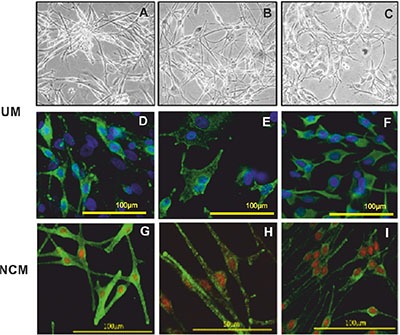
Brightfield images of UM cells with a spindle phenotype (A and B) and plump epithelioid-like cells (C). Immunofluorescence phenotyping of UM (D, E and F) and NCM (G, H and I) cells in culture. UM cells expressing (D) MelanA, (E) HMB45 and (F) vimentin. Positive staining was detected with Alexa 488 (green) and nuclei were counterstained with DAPI (blue). NCM expressing (G) MelanA, (H) gp100 and (I) HMB45. Positive staining was detected with Alexa 488 (green) and nuclei were counterstained with propidium iodide (red).