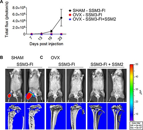
Figure 8. Bone preconditioning by SSM2 cells is not sufficient to allow estrogen-independent growth of SSM3 cells.

(A–C) 105 SSM2 and 105 SSM3-Fl cells were co-injected into the right tibia of WT female mice, one week after OVX surgery (n = 6). SHAM-operated and OVX mice were injected with 105 SSM3-Fl cells into the right tibia alone and used as controls (n = 6/group). (A) Bioluminescence imaging monitoring SSM3-Fl tumor growth (mean +/− sem) in SHAM-operated mice (black circles) and OVX mice (red squares for SSM3-Fl alone and blue triangles for SSM3-Fl + SSM2 cell co-injection). (B, C) 2 out of 6 representative bioluminescence images and viva-CT scans of the right tibias of SHAM-operated (B) and OVX (C) mice from (A) at 27 days post tumor injection are shown. Osteolytic lesions are depicted by arrows.
