
Figure 5. DHA-rich environment retards tumor growth in vitro.
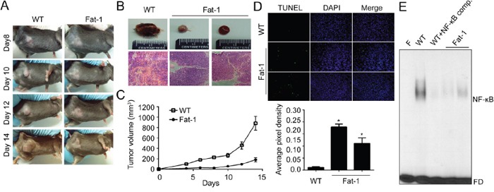
A-B. Fat-1 transgenic and C57BL/6 wild-type mice were implanted with murine breast carcinoma cell line EO771 cells. After inoculation, animals were closely monitored for the development of subcutaneous tumor. After two weeks, the animals were sacrificed and xenograft tissues were collected for further experiment. Day of the implantation of the tumor cells was designated day 0. WT, representative xenograft tumor from wild type mouse; Fat-1, two representative xenograft tumors from fat-1 transgenic mice. C. The tumor size was measured at indicated time with a caliper. Tumor volume=0.5×(width)2×length. D. The animals were sacrificed at two weeks after implanted, tumor tissues were fixed with formalin and TUNEL stain was performed. E. The nuclear extracts were prepared from mice xenograft tissues. Then the nuclear extracts were incubated with the radiolabeled oligonucleotide containing NF-κB and EMSA was performed. F, blank without nuclear extract; Comp., competitor without isotope labeling; FD, free DNA probe.