
Figure 1. Mammary carcinoma cells exchange vesicles with nontransformed mammary fibroblasts.
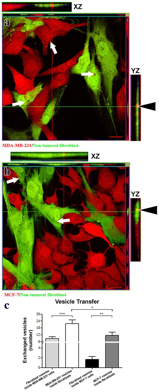
Panels a. and b. present confocal images showing exchange of vesicular structures between fibroblasts (green) and tumor cells (red, arrows in panels a and b). Orthogonal projections verify the presence of tumor vesicles inside fibroblasts (arrowheads in panels a and b). Panel c. presents quantitation of vesicle transfer as numbers of vesicles transferred in a field containing 20 cells (mean+SEM, n≥5). Single, double, and triple asterisks indicate statistically significant differences of p<0.05, p<0.01 and p<0.001, respectively. Scale bars = 20 μm.