
Figure 4. AHNAK localization in cell protrusions and extracellular vesicles.
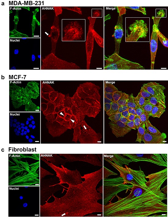
AHNAK was visualized in cells by immunofluorescence confocal microscopy. a. MDA-MB-231 cells contained AHNAK at cell edges, as dot-like structures in the lamellipodia (insert), and in released vesicles (arrow). b. MCF-7 cells contained AHNAK mostly at intercellular junctions (arrowheads) and vesicles (arrows). c. Nontransformed fibroblasts contained AHNAK at cell edges and as small dots throughout the cytoplasm. Scale bars = 5 μm.