
Figure 9. AHNAK is highly expressed in human mammary carcinoma cells in vivo.
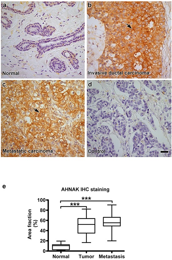
AHNAK expression was evaluated in human clinical samples by immunohistochemistry. a. normal mammary epithelium, b. invasive ductal carcinoma, c. metastatic carcinoma from breast cancer patients, and d. invasive ductal carcinoma without primary antibody as a negative control. e. Quantitation of these data is shown as the percent of area with AHNAK expression (mean±SEM), and indicate that AHNAK expression was increased in invasive ductal carcinoma (n=35) and metastatic carcinoma (n=10) compared to normal tissue (n=9). Triple asterisks indicate significant differences compared to normal tissue with p<0.001. Scale bar = 20 μm.