
Figure 4. Colorectal cancer exosomes increase the expression of vacuolar H+-ATPase (V-ATPase) and CEA in colonic (c) MSCs and colon cancer (cc) MSCs.
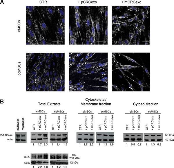
(A) Confocal laser scanning microscopy of 5 cMSC cells optical sections Z-projection taken from the bottom to the edge of cMSCs treated with primary CRC exosomes (pCRCexo) or with metastatic CRC exosomes (mCRCexo) for 72 hours and incubated with primary anti-V-ATPase antibody, followed by Alexa Fluor®-488-conjugated secondary Ab (shown in white). Nuclei are reported in blue (DAPI). Scale bars, 40 μM. (B) Western blot analyses of V-ATPase, CEA and actin proteins, performed in total protein extracts of: pCRCexo or mCRCexo (50 μg), cMSC or ccMSC cells treated with pCRCexo or mCRCexo; Western blot analyses of V-ATPase and actin in Cytoskeletal/Membrane and Cytosol fractions; untreated cells (CTR). Results of densitometry analyses are reported as fold-increase in the expression of each molecule, related to actin loading.