
Figure 3. Endogenous phosphorylated p21 and p21S130D are less stable.
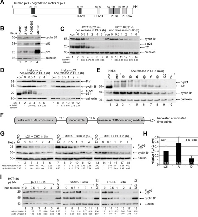
A. Schematic illustration of human p21 including its different known destruction motifs (gray boxes) like F-box, D-box, caspase cleavage site (DHVD), PEST sequence and PIP-box. B. HeLa cells were treated overnight with the pan-caspase inhibitor Z-VAD, the calpain inhibitor PD150606 and the proteasome inhibitor MG132 for Western blot analyses with indicated antibodies. DMSO served as vehicle and calnexin as loading control. C. HCT116 p21+/+ and p21−/− cells were synchronized to prometaphase with nocodazole (noc), released into CHX-containing medium for indicated time and harvested for Western blot analysis. Calnexin served as loading control. Ratio of cyclin B1/calnexin is shown. D. HeLa cells transfected with control siRNA (sicon) or siRNA against p21 (sip21) were synchronized to prometaphase with nocodazole (noc). The cells were released into CHX-containing medium. Calnexin served as loading control. Ratio of cyclin B1/calnexin is shown. E. HeLa cells were synchronized to prometaphase with nocodazole (noc), released into medium containing CHX for indicated time points for Western blot analysis. Non-treated or nocodazole treated cells were taken as controls. Calnexin served as loading control. F. Working schedule of the CHX kinetics. G. HeLa cells were transfected with different p21-constructs, synchronized with nocodazole and released in CHX-containing medium. Cells treated with the pan-caspase inhibitor Z-VAD were taken as control. Tubulin served as loading control. H. Quantification of the FLAG level normalized against the loading control is shown as mean ± SEM from three independent experiments. Time point: 4 h after CHX treatment. I. CHX release kinetics was also performed with HCT116 p21−/− cells expressing FLAG-p21, S130A or S130D. MG132 treated cells served as positive and β-actin as loading control.