
Figure 5. Interfering with phosphorylation of S130 in p21 extends mitotic duration.
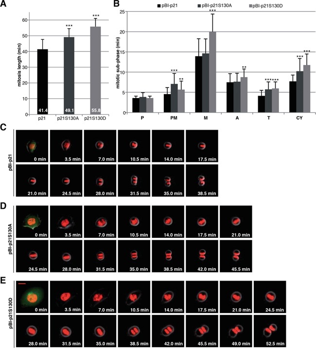
A. HCT116 p21−/− cells stably marked with H2B-tdTomato and transfected with different pBI-p21 constructs were subjected to time-lapse imaging. pBI vector is a mammalian bidirectional expression vector designed to express a protein of interest and a green fluorescent protein (ZsGreen) as transfection marker shown at 0 min. The duration of mitosis, captured by time-lapse imaging at 3.5 min image intervals, was evaluated (n=50 cells of each condition). The results are presented as mean ± SD and statistically analyzed. ***p < 0.001. B. The time of each mitotic subphase was evaluated (n=50 cells per each cell line). The results are presented as mean ± SD and statistically analyzed between wild type pBI-p21 and pBI-p21S130A/D. **p < 0.01, ***p < 0.001. P: prophase, PM: prometaphase, M: metaphase, A: anaphase, T: telophase, CY: cytokinesis. C-E. Representative pictures of mitotic cells from pBI-p21 (C), pBI-p21S130A (D) and pBI-p21S130D (E) are shown. Scale bar: 20 μm.