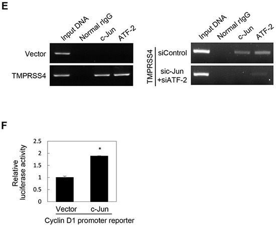
Figure 2. JNK signaling activity and c-Jun/ATF-2 were required for TMPRSS4-mediated Slug and cyclin D1 induction.

A. PC3 cells were transfected with a TMPRSS4 expression vector for 24 h and then treated with pharmacological inhibitors for 24 h before whole-cell lysates were prepared for immunoblotting as described in the Materials and Methods. B. PC3 cells were co-transfected with a TMPRSS4 expression vector and an AP-1 reporter plasmid for 48 h. AP-1 activity was determined by a reporter assay as in Figure 1D. C. PC3 cells were co-transfected with a TMPRSS4 expression vector or an empty vector and siRNA specific to c-Jun or ATF-2 or negative control siRNA for 48 h. Transfected cells were lysed and used for immunoblotting. An anti-myc antibody was used to detect myc-tagged TMPRSS4. GAPDH was used as an internal control. D. Left: PC3 cells were co-transfected with a c-Jun expression vector and a Slug promoter (−981/+174) reporter construct for 48 h. Reporter assays were performed as in Figure 1D. Right: PC3 cells were transfected with a c-Jun expression vector for 48 h and lysed for immunoblotting. E. ChIP analysis of the interaction of c-Jun and ATF-2 with the Slug promoter. Left: Chromatin fragments from PC3 cells transfected with a TMPRSS4 expression vector or an empty vector for 48 h were immunoprecipitated with control normal rabbit IgG, anti-c-Jun, or anti-ATF-2 and analyzed via PCR using Slug promoter primers (+1/+172). The input control is in lane 1. Right: ChIP assay using PC3 cells co-transfected with a TMPRSS4 expression vector and siRNAs specific to c-Jun or ATF-2 for 48 h. F. PC3 cells were co-transfected with a c-Jun expression vector and a cyclin D1 promoter (−962/+134) reporter construct for 48 h. Reporter assays were performed as in Figure 1D. Values represent mean ± SD. * P < 0.05.
