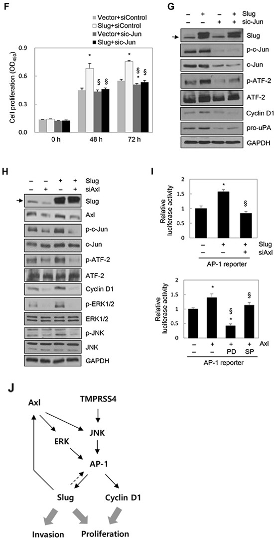
Figure 4. Slug activated AP-1 and induced cyclin D1, leading to cell proliferation.

A. PC3 cells were transfected with a Slug expression vector for 48 h. Transfected cells were lysed and used for immunoblotting. Conditioned medium was collected after an additional 48 h and analyzed by immunoblotting (pro-uPA and active uPA). An anti-myc antibody was used to detect myc-tagged Slug. B. Cells were co-transfected with a Slug expression vector and an AP-1 reporter plasmid for 48 h. Reporter assays were performed as in Figure 1D. Values represent mean ± SD. * P < 0.05. C. HEK293E cells were transfected with a Slug expression vector for 48 h. Transfected cells were lysed and used for immunoblotting. β-Actin was used as an internal control. D, E. PC3 cells were transfected with siRNA specific to Slug for 48 h. (D) Transfected cells were seeded into 96-well plates at a density of 3000 cells/well and incubated for 48 or 72 h. Cell proliferation was determined by the colorimetric WST assay. Values represent mean ± SD. * P < 0.05. (E) Transfected cells were lysed and used for immunoblotting. F, G. PC3 cells were co-transfected with a Slug expression vector or an empty vector and siRNA specific to c-Jun or negative control siRNA for 48 h. (F) Transfected cells were seeded into 96-well plates at a density of 3000 cells/well and incubated for 48 or 72 h. Cell proliferation was determined by the colorimetric WST assay. Values represent mean ± SD. * P < 0.05 compared with empty vector + control siRNA; § P < 0.05 compared with Slug + control siRNA. (G) Transfected cells were lysed and used for immunoblotting. Arrow indicates endogenous Slug (C, G, H). H. PC3 cells were co-transfected with a Slug expression vector and Axl-specific siRNA for 48 h prior to lysis for immunoblot analysis. I. Upper: PC3 cells were co-transfected with a Slug expression vector, an AP-1 reporter plasmid, and Axl-specific siRNA for 48 h. Lower: PC3 cells were co-transfected with an Axl expression vector and an AP-1 reporter plasmid for 6 h and then treated with pharmacological inhibitors for 24 h. (Enhanced expression of Axl by the Axl expression vector was confirmed by immunoblotting (Supplementary Figure S3E)). Reporter assays were performed as in Figure 1D. Values represent mean ± SD. * P < 0.05 compared with empty vector + control siRNA (upper) or empty vector + DMSO (lower); § P < 0.05 compared with Slug + control siRNA (upper) or Axl + DMSO (lower). J. A schematic representation of the pathway for TMPRSS4-induced invasion and proliferation in human cancer cells. Axl is involved in Slug-mediated AP-1 activation.
