
Figure 7. Depletion of CBS decreases cell proliferation and migration.
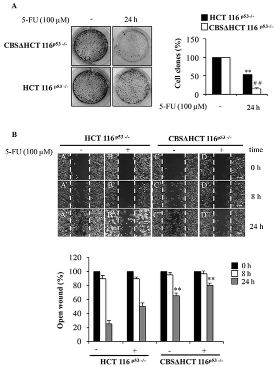
A. Representative images of clonogenic analysis for cell proliferation in HCT 116p53−/− and CBSΔHCT 116p53−/− cells after 5-FU treatment. **P < 0.01 vs. untreated HCT 116p53−/− cells; ##P < 0.01 vs. untreated rpL3Δ HCT 116p53−/−cells. B. Representative images of wound healing assay for cell proliferation in HCT 116p53−/− and CBSΔHCT 116p53−/− cells after 5-FU treatment. Wound widths were measured at 0, 8 and 24 h on 3 fields per well and averaged. Data is expressed as the fold-decrease of area respect to controls (A, B, C, D) set as 100%. **P < 0.01 vs untreated HCT 116p53−/− cells.