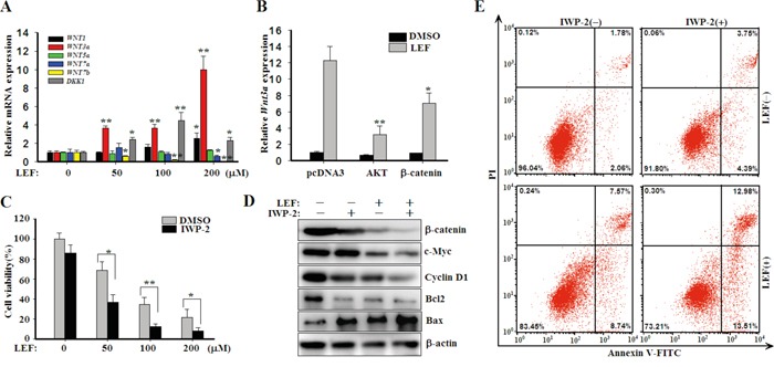
Figure 6. LEF upregulates WNT ligands to compromise cytotoxic effects.

A. Real-time PCR for the expression of WNT1, WNT3a, WNT5a, WNT7a, WNT7b, and DKK1 in mRNA levels. Data represent mean ± SD from three independent experiments. B. Caki-2 cells were transfected with plasmids encoding AKT or β-catenin as depicted, and then cells were treated with 200 μM LEF for 48 h to detect the expression of WNT3a mRNA by real-time PCR. C. Cell viability was estimated by MST assay after Caki-2 acells were incubated with increasing concentrations of LEF together with 20 μM IWP-2 for 48 h. All experiments were done in triplicates and each bar represents mean ± SD (*P<0.01, ** P<0.05, vs. the control). D. Changes of growth and apoptosis-associated proteins after combined treatment of LEF and IWP-2 for 48 h. Representative images from at least three independent experiments are shown. E. Flow cytometry analysis of apoptosis was determined in Caki-2 cells treated with 200 μM LEF and 20 μM IWP-2 for 48 h. Data are typical of three similar experiments. The percentage of Annexin V-FITC and/or PI positive cells was depicted with cytofluorometer quadrant graphs.
