
Figure 4. HIFU exposure suppressed melanoma metastasis and down-regulated miR-21 expression in vivo.
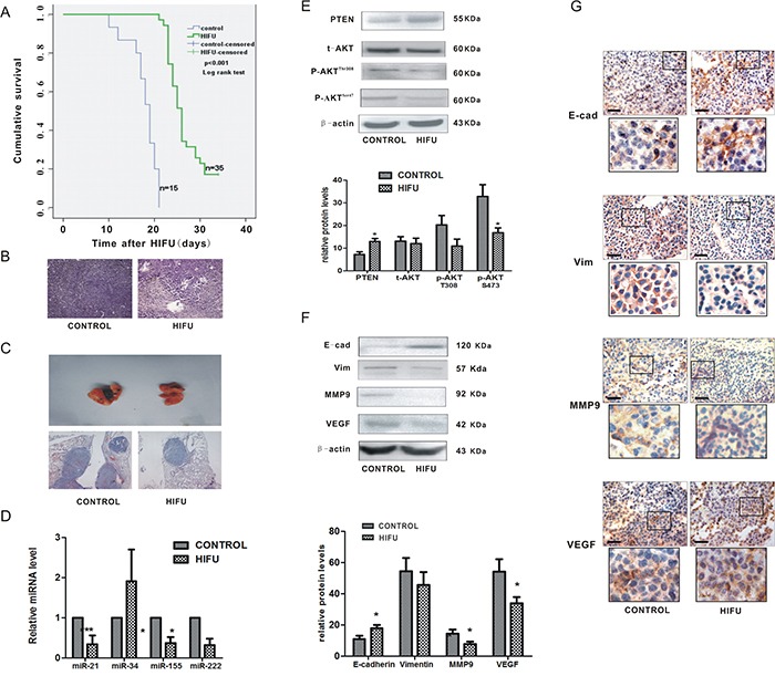
A. The cumulative survival curves in both groups using Kaplan-Meier analysis. B. Representative images of histological tissue sections of mice 14 days after HIFU or sham-HIFU exposure. Magnification, ×100. C. Representative H-E staining images of lungs of mice after subcutaneous inoculation of B16-F10 melanoma cells and treated with HIFU or sham-HIFU are shown in the upper panel. ×100. D. The alteration of microRNAs relative expression was detected by qPCR in residual tumor tissues after HIFU exposure. ***p<0.001, *p<0.05. E. The expression of PTEN, AKT and p-AKT in the residual tissues after HIFU exposure were detected using Western blot analysis in the upper panel. β-actin was used as an internal reference control. The quantified relative expression of proteins were showed in the lower panel.*p<0.05. F. The proteins in residual tumor tissues were detected by Western blot. β-actin was detected as an input control. The densitometric ratios are shown in the lower panel.*p<0.05. G. Immunohistochemical analysis was used to investigate the relative expression of proteins involved in metastasis. Scale bar, 100 um.