
Fig. 2. Preferential ERK phosphorylation in the mPFC after systemic administration of METH versus cocaine.
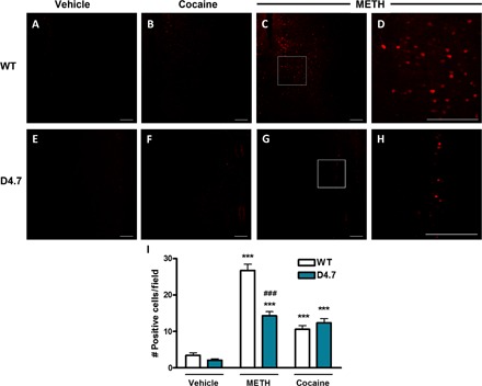
(A) to (C) and (E) to (G) Immunohistochemical analysis of phosphorylated ERK1/2 in the mPFC of WT (upper row) or D4.7 (lower row) mice treated with vehicle (A and E), cocaine (30 mg/kg; B and F), or METH (3 mg/kg; C and G). (D and H) Magnification of the framed area in (C) and (G), respectively. Scale bars, 200 μm. (I) Quantification of positive cells from 400-μm × 400-μm fields in the mPFC. Values are means ± SEM of 32 to 57 different fields obtained from three different animals per condition. Statistical significance was calculated by one-way ANOVA followed by Bonferroni’s post hoc tests (***P < 0.001, compared with the respective vehicle, and ###P < 0.001 compared with WT).