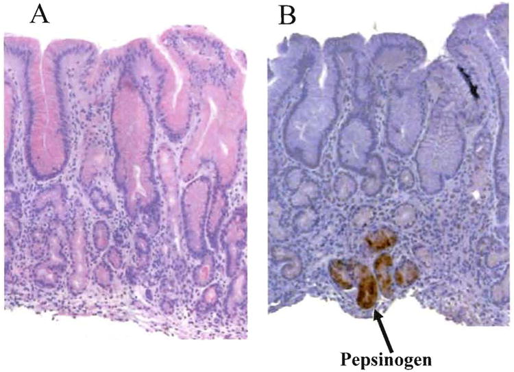
Figure 3. Pseudopyloric metaplasia.

Corpus biopsies with pseudopyloric metaplasia. A shows mucosa that without knowledge of its source as corpus mucosa would likely be identified as antrum by the pathologist. B. Immunohistochemical staining for pepsinogen I showing positive staining confirming that the tissue is actually atrophic corpus mucosa (courtesy of the Gastrointestinal mucosal pathology laboratory, Baylor College of Medicine, Hala El-Zimaity director).
