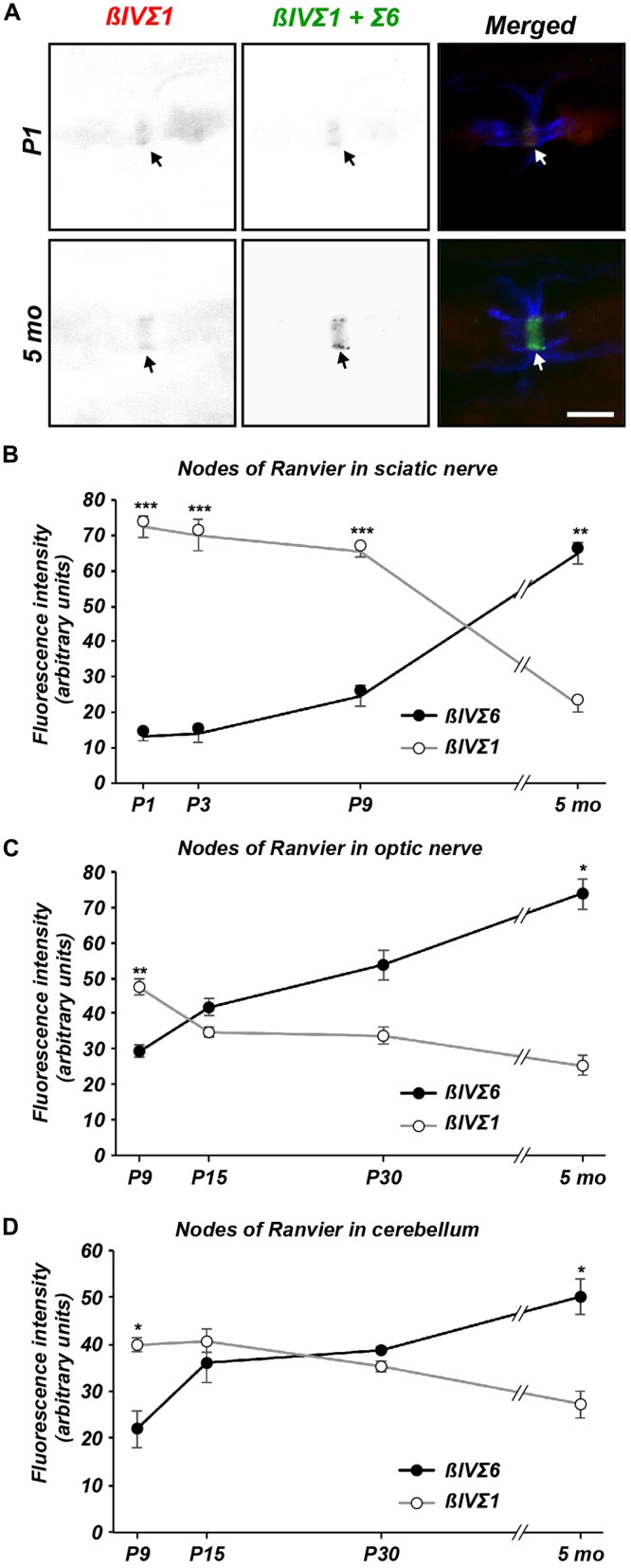
Figure 4.

Differential expression of βIV spectrin splice variants at nodes of Ranvier. (A) Immunolabeling of nodes of Ranvier using NT (βIVΣ1) and SD (βIVΣ1 + βIVΣ6) antibodies in P1 and 5 sciatic nerve. Caspr immunostaining is shown in blue. Scale bar = 5 μm. (B–D) Quantification of relative βIVΣ1 and βIVΣ6 expression in sciatic nerve (B), optic nerve (C) and cerebellar nodes (D) as a function of age. Error bars ± SEM. *p < 0.01, **p < 0.001, ***p < 0.0001.
