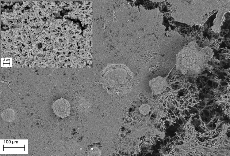
Fig 4. S. mutans ATCC25175 biofilm (plaque) formed on the surface of bone (Magn.275X).
The dense structure of plaque is seen in the right and central part of the image, whereas plaque disruption is visible in the right portion of the image revealing multilayer composition and biofilm matrix (upper left inset, Magn. 660X). Media: artificial saliva plus 3% sucrose. SEM Zeiss EVO MA25 microscope.

