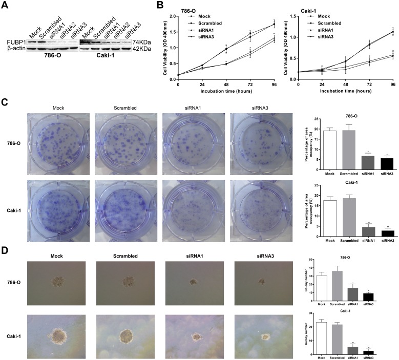
Fig 2. FUBP1 depletion decreases cell proliferation of 786-O and caki-1.
(A) FUBP1 was knocked down in 786-O and caki-1 cells, and FUBP1 protein levels were monitored by Western blot analysis with the anti-FUPB1 antibody. β-actin was used as the loading control. (B) Assessment of 786-O and caki-1 cell proliferation by MTS assays. (C) Colony formation assays were performed to determine the proliferation of 786-O siRNA1/3, caki-1 siRNA1/3, and the corresponding control cells (786-O-Mock/Scrambled and caki-1-Mock/Scrambled). The area percentage occupied by 786-O-siRNA1/3 and caki-1-siRNA1/3 cells was markedly less than those of 786-O-Mock/Scrambled and caki-1-Mock/Scrambled. (D) Representative images of soft agar colony assay in cells transfected with indicated siRNAs. Original magnification: 100×. Colony count statistics showed a significant reduction in siRNA1/3 786-O and caki-1 cells. Values are expressed as the mean ± SD of three independent experiments, *P < 0.05, **P < 0.01.

