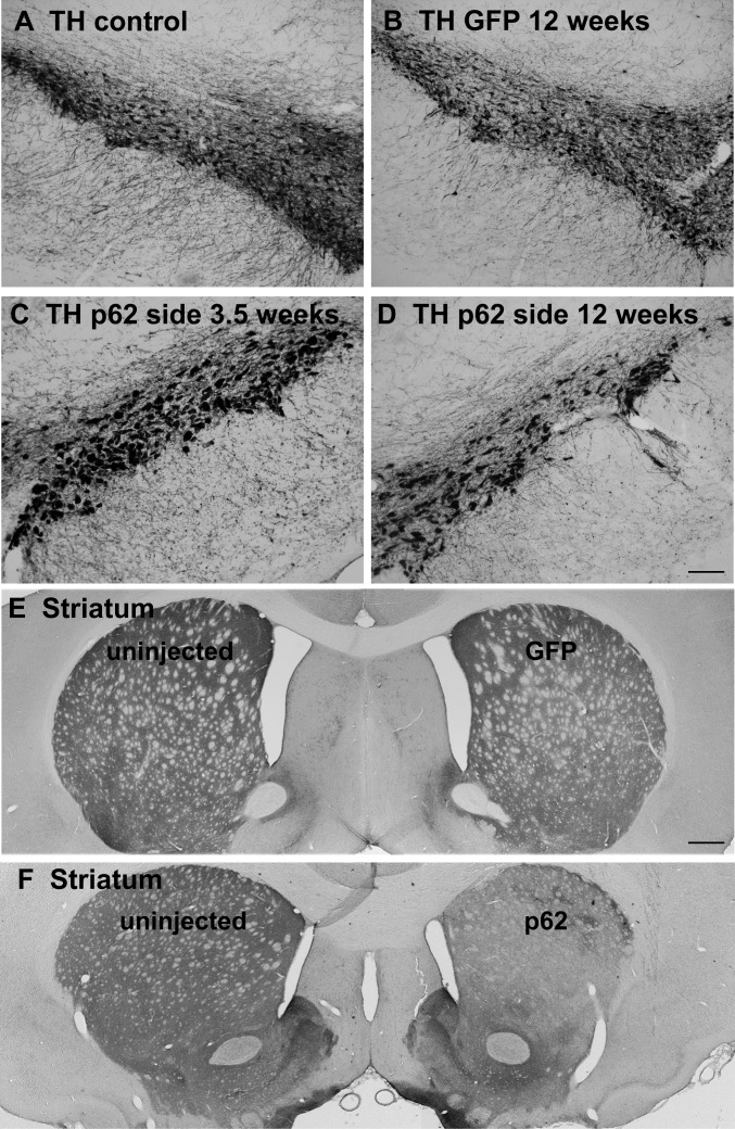
Fig 1. Tyrosine hydroxylase staining of dopaminergic neurons in the nigrostriatal pathway.
A) Substantia nigra from an uninjected side of the brain. B) Substantia nigra from an AAV9 GFP injected side after 12 weeks after gene transfer. C) Substantia nigra from an AAV9 p62 injected side at 3.5 weeks after gene transfer. D) Substantia nigra from an AAV9 p62 injected side at 12 weeks after gene transfer. There is a noticeable hypertrophy of the neurons on the p62 side at both intervals yet an apparently progressive loss of cells between the two time points in the p62 group. E) Forebrain from a GFP animal at 12 weeks after gene transfer. F) Forebrain from a p62 animal at 12 weeks after gene transfer. There is a loss of striatal tyrosine hydroxylase on the side where AAV9 p62 was injected into the substantia nigra. Bar in D = 134 μm; same magnification in A-C. Bar in E = 536 μm; same magnification in F.

