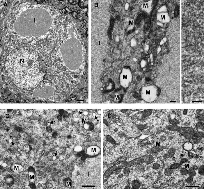
Fig 5. Electron microscopy: filamentous inclusions and disruption of mitochondria induced by p62 gene transfer.
A, B) Consistent with the p62-positive inclusions observed by light microscopy, there were numerous inclusions (I) viewed within neurons on the AAV9 p62 injected side, but not found on the uninjected side. The filaments were densely packed in non-membrane bound inclusions and had a width of approximately 10 nm (right panel in B). B, C) In cells with the inclusions, the mitochondria (M) were grossly abnormal with the disruption of cristae structure and the formation of vacuoles within the mitochondria. Arrows point to small vesicles. D) A sample from the uninjected side of the brain shows normal mitochondria. Time point of 9 days after gene transfer in A, and 21 days after gene transfer in B-D. Bar in A = 1 μm; bar in B left = 0.2 μm, bar in B right = 50 nm; bar in C, D = 0.1 μm.

