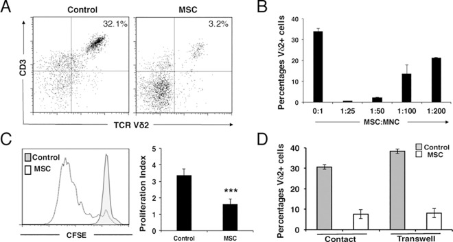
Fig 1. MSCs inhibit the expansion of Vδ2+ cells by soluble mediators.
(A) The presence of MSCs reduces the expansion of Vδ2+ cells. Representative flow cytometric analysis of Vδ2+ cells activated from whole PBMC by HDMAPP and rh-IL2 for seven days in the absence (left panel) or presence (right panel) of MSCs. (B) Increasing ratios of MSC:MNC diminish the inhibitory effects of MSCs on Vδ2+ cell proliferation. Results show the means ± S.D. of triplicate samples. (C) Total PBMCs were labeled with CFSE and activated by HDMAPP and rh-IL2. Analysis of Vδ2+ cell proliferation in the presence (white) or absence (grey) of MSCs was performed after five days by Flow Cytometry. The presence of MSCs lowers the proliferation index of Vδ2+ cells (right panel). Results show the means ± S.D. of triplicate samples. ***P ≤ 0.001. (D) Analysis of the percentage of Vδ2+ cells cultured in cell-to-cell contact or in a transwell system in the presence/absence of MSCs. Vδ2+ cell expansion is inhibited in the same way in both systems indicating that soluble factors are responsible for immunoregulation. Results show the means ± S.D. of triplicate samples.

