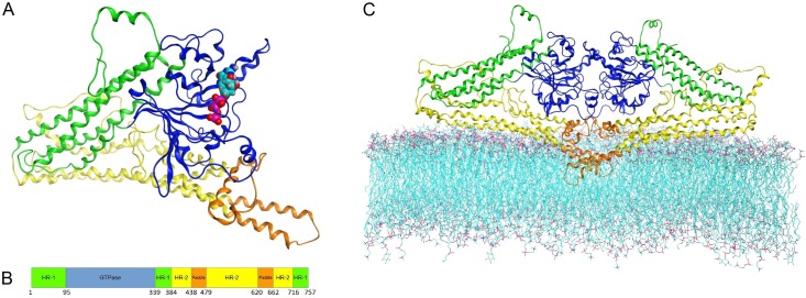
Fig 6. Overall MFN2 model structure.
Colors demonstrate domain division: GTPase (blue), HR-1 (green), HR-2 (yellow), and paddle region (orange). A. MFN2 monomer structure with visible GTP showed as Van der Waals spheres. B. Sequence-oriented MFN2 domain composition, with boundary residues. C. MFN2 homodimer embedded in lipid bilayer. Paddle residues 620–662 deeply embedded, residues 438–479 localized above hydrophobic layer.

