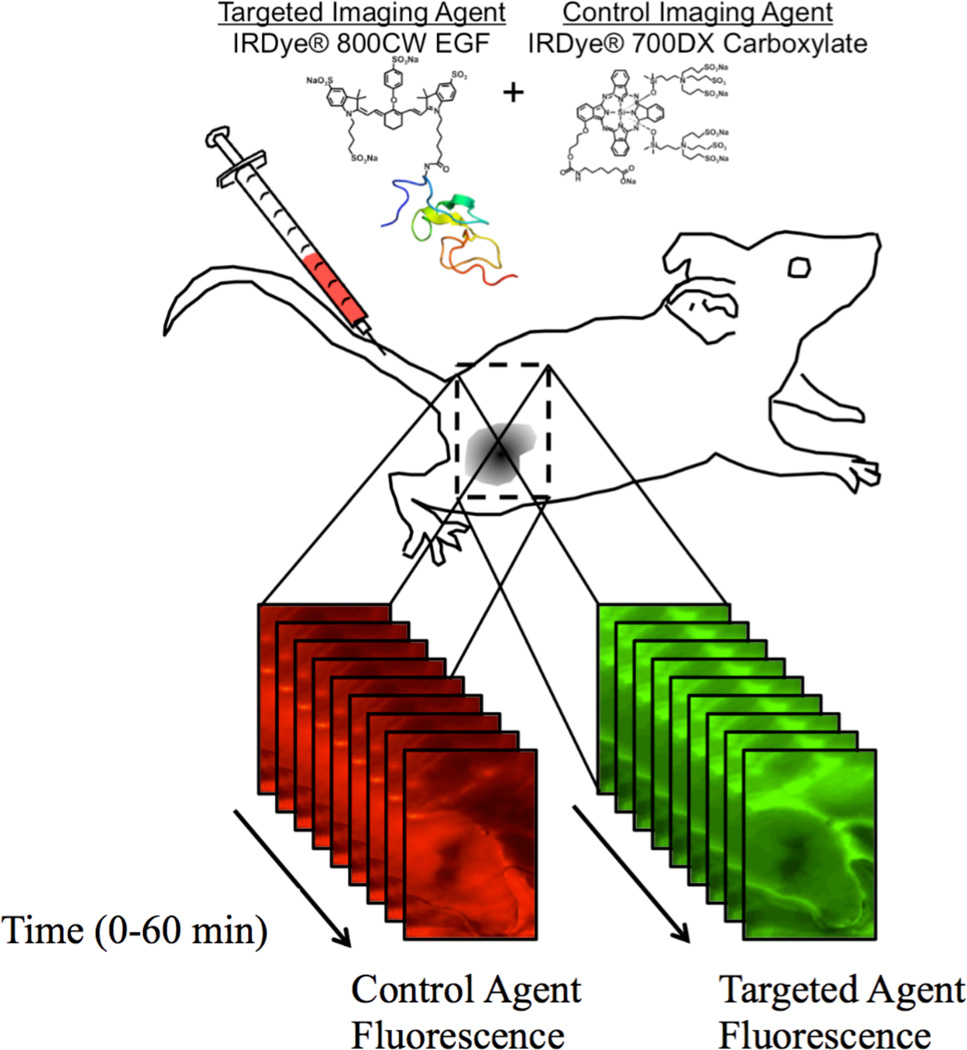
Figure 2.
In the current study, the targeted imaging agent was fluorescently labeled epidermal growth factor (IRDye® 800CW EGF) and the control imaging agent was an unbound fluorescent molecular (IRDye® 700DX Carboxylate). Temporal kinetics of both imaging agents were measured in surgically exposed subcutaneous xenograft tumors grown in severe combined immunodeficiency (SCID) mice using a dual-channel fluorescent scanner (Odyssey® Imaging System, LI-COR Biosciences) at 2–5-min intervals up to 60 min post-agent-injection.

