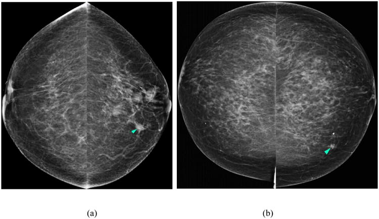
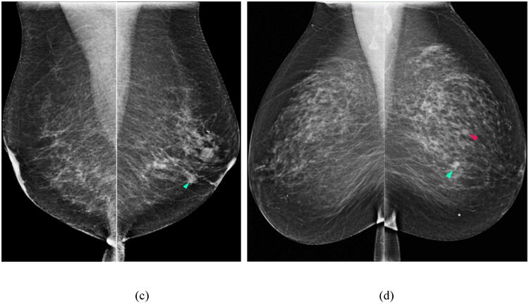
Figure 1.


Two examples of applying our lesion based computer-aided detection (CAD) scheme on two cancer cases in our image dataset. Figures 1 (a) and (c) display the first example with a cancerous mass detected by the scheme (i.e., indicated with light blue arrow) in both the CC and MLO images of the left breast. Figures 1 (b) and (d) display the results obtained on the second example – a cancerous mass was marked with a FP detection (magenta arrow).
