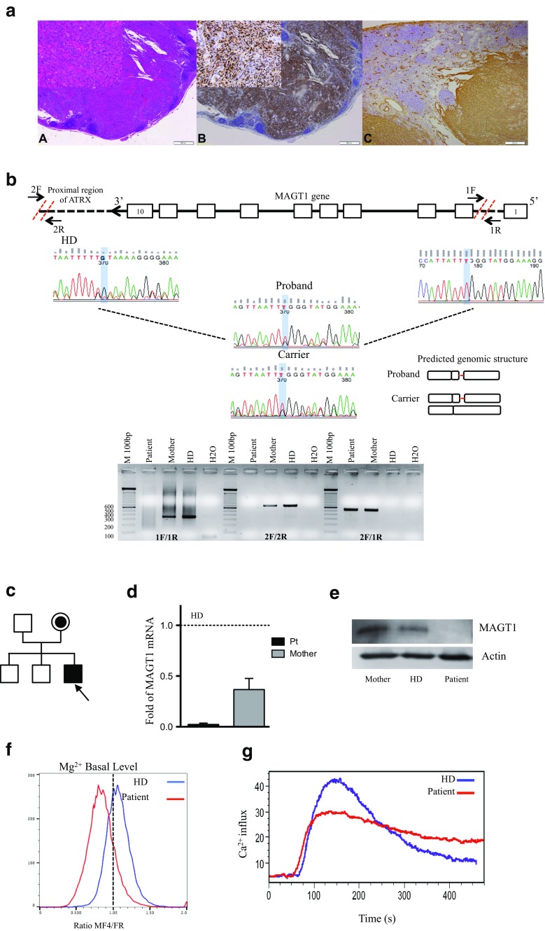
Fig. 1.
Characterization of X-MEN patient. a Lymph node involvement by Kaposi sarcoma: A diffuse proliferation of spindle cells forming slits containing red blood cells (HE, 4×, inset 20×), B HHV-8 positivity in nuclei of spindle cells (4×, inset 20×), and C CD34 positivity of spindle cells (10×). b Schematic representation of primers’ design for the identification of the deletion in MAGT1 gene. Deletion in the proband and carrier was confirmed by genomic amplification/DNA sequencing. Predicted structure is showed. c Pedigree of the family. The patient is indicated by an arrow. d Quantitative RT-PCR showing expression of MAGT1 mRNA in T cells normalized to telomerase. Results represented as relative to HD. e Western blot on T cells from patient, mother, and HD. MAGT1 30KDa. Actinβ 40 kDa. f Mg2+ basal levels in the patient (red line) and HD (blue line). g Calcium flux in freshly isolated PBMC from the patient and HD stimulated with anti-OKT3 (5 μg/mL) shown as percentage of responding cells as a function of time. HD: healthy control

