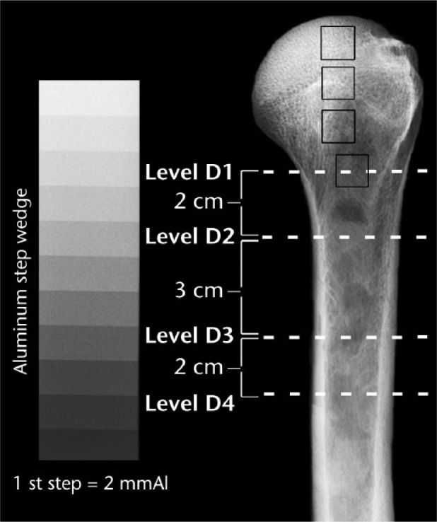
Fig. 1.

Anteroposterior view of a left cadaver humerus showing the four measurement locations (dashed white lines) of the metaphysis/diaphysis, where D1 is the surgical neck. From top to bottom, the dark squares indicate head (H)1, H2, H3 and D1 locations where mmAl measurements were made.
