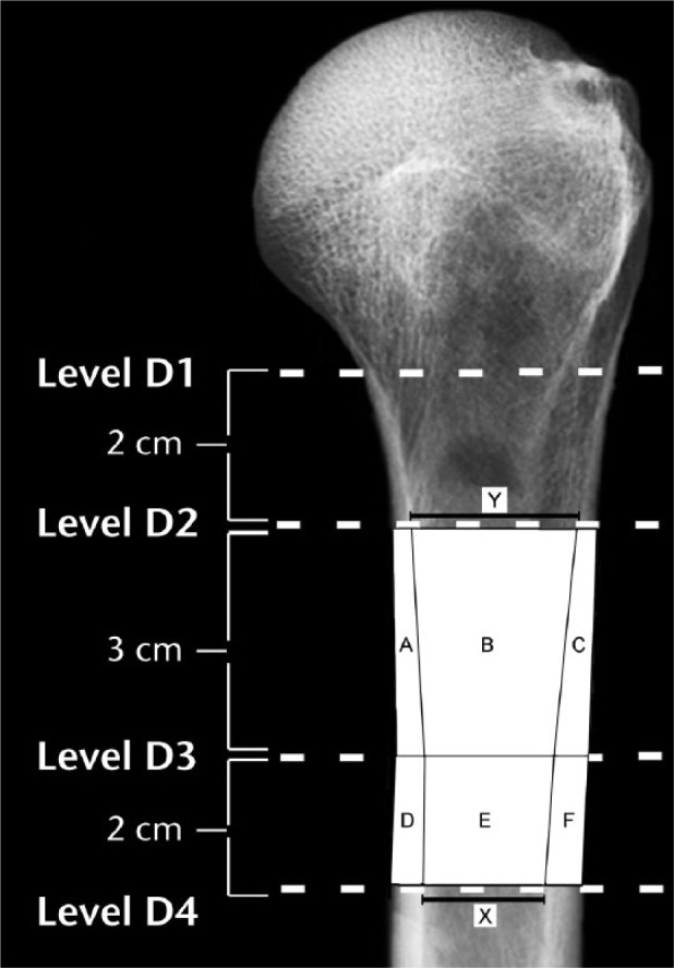
Fig. 2.

In terms of the areas A-F that are shown diagrammatically in this radiograph, areal cortical index for the D2-D3 region is: [((A + B + C) – B) / (A + B + C)]. Areal cortical index for the D3-D4 region is: [((D + E + F) – E) / (D + E + F)]. The canal-to-calcar ratio is calculated as X/Y; as shown, these values were obtained from the D2 and D4 locations. The medial cortical ratio and an example of a linear cortical index measurement are shown in Figure 3. (D2, 2 cm below surgical neck; D3, 5 cm below surgical neck; D4, 7 cm below surgical neck.)
