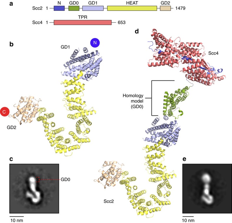
Figure 1. Structure of Scc2 hook and the full-length Scc2–Scc4 model.
(a) Schematics showing the linear domain organizations of Scc2 and Scc4. The same colouring scheme is used in the corresponding crystal structures of Scc2 and Scc4 as shown in (b,d). (b) The modular structure of Scc2 resembles a ‘hook'. The overall fold comprises an N-terminal globular domain (GD1; violet), 14 contiguous HEAT repeats (yellow) and an oval-shaped C-terminal globular domain (GD2; wheat) with an extended loop connecting the C-terminal capping helix. (c) An EM 2D class average of Scc2 hook with GD0 indicated. (d) Reconstruction of a pseudo-full-length structure of Scc2–Scc4 with the previously determined crystal structure of Scc21–168–Scc434–620 (Scc2N–Scc4, PDB I.D. 5C6G; purple and salmon), a homology fold of Scc2169–377 (GD0; mauve) from human symplekin (PDB I.D. 3O2T) and Scc2 hook (violet, yellow and wheat). (e) An EM 2D class average of full-length Scc2–Scc4.

