. 2016 Sep 30;49(9):502–507. doi: 10.5483/BMBRep.2016.49.9.030

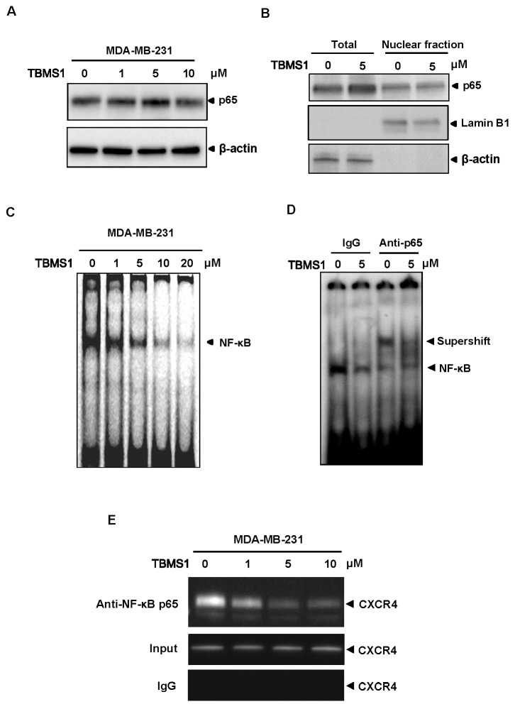
Fig. 2. TBMS1 suppresses the binding of NF-κB to CXCR4 promoter. (A) MDA-MB-231 cells were treated with TBMS1 for 24hr as indicated concentrations, and the expression of p65 and β-actin were analyzed by immunoblotting. (B) MDA-MB-231 cells were treated with 5 μM TBMS1 for 24 hr. The total cell lysate and the nuclear extracts were analyzed with the indicated antibodies by immunoblotting. The nuclear envelope protein Lamin B1 was used as a nuclear marker. (C) MDA-MB-231 cells were treated with TBMS1 for 24 hr at indicated concentrations. The nuclear extracts were used in EMSA assay with the labeled CXCR4 promoter sequence containing NF-κB binding site. (D) MDA-MB-231 cells were treated with 5 μM TBMS1 for 24 hr. The nuclear extracts were incubated with the labeled CXCR4 promoter sequence containing NF-κB binding site in the presence of 1 μg IgG control or anti-p65 antibody, as indicated. The NF-κB binding activity was analyzed by EMSA assay. Specific shift bands for NF-κB were indicated with an arrow. (E) MDA-MB-231 cells were pretreated with TBMS1 for 24 hr and the proteins were cross-linked with DNA with formaldehyde, and then subjected to ChIP assay with an anti-p65 antibody. PCR on the immunoprecipitate was performed using primers spanning -192 to +45 of the CXCR4 promoter. Reaction products were resolved by electrophoresis. Representative results of three independent experiments are shown.

Fig. 2.