. 2016 Sep 30;49(9):502–507. doi: 10.5483/BMBRep.2016.49.9.030

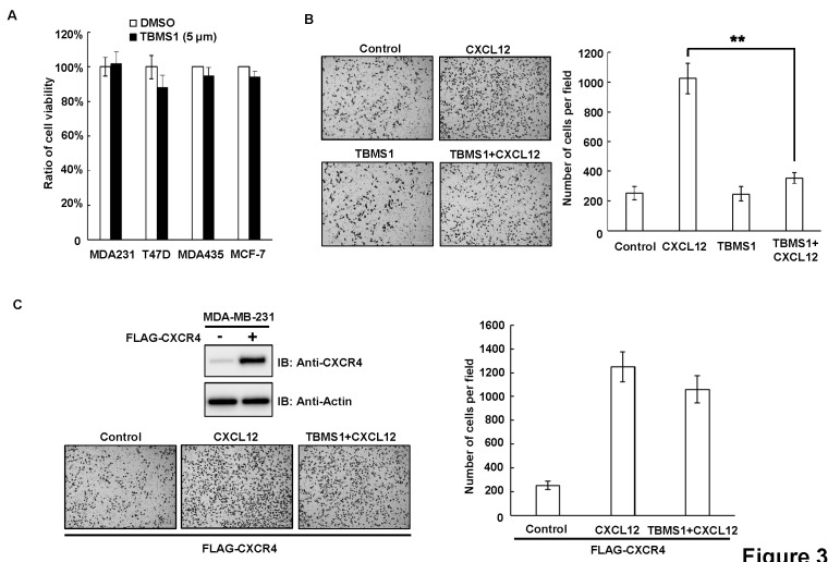
Fig. 3. TBMS1 suppresses the CXCL12-induced invasion of breast cancer cells. (A) MDA-MB-231, T47D, MDA-MB-435 and MCF-7 cells were treated with DMSO or TBMS1 (5 μM) for 24 hr. Relative cell viability was analyzed by MTT assay. Results shown are averages ± S.E.M. (B) MDA-MB-231 cells were treated with/out CXCL12 (300 ng/ml) combined with/out TBMS1 (5 μM) as indicated, for 24 hours. Cell invasion was analyzed by Tranwell assay. Left, representative images of Transwell assay. Right, quantitative data of migrated cells. Results shown are averages ± S.E.M. (C) MDA-MB-231 were transfected with FLAG-CXCR4 vector. Upper-left, expression levels of CXCR4 were examined by immunoblotting in empty vector and pCMV6-CXCR4 transfected MDA-MB-231 cells. Lower-left, representative images of transwell assay. Right, quantitative data of migrated cells. Results shown are averages ± S.E.M.

Fig. 3.