. 2016 Sep 30;49(9):520–525. doi: 10.5483/BMBRep.2016.49.9.098

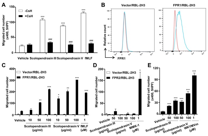
Fig. 3. Neutrophil chemotaxis stimulated by the two AMPs is mediated by FPR1. (A) Mouse neutrophils were incubated in the absence or presence of CsH (10 μM) for 30 min, and were applied to the upper well of a multiwell chamber containing 100 μg/ml of the two AMPs or 1 μM of fMLF for 90 min. (B) Cell surface expression of FPR1 was determined by flow cytometric analysis (red, isotype control; blue, anti-FPR1). The results are representative of three independent experiments. (C) Vector- or FPR1-expressing RBL-2H3 cells were applied to the upper well of a multiwell chamber containing several concentrations (0 μg/ml, 10 μg/ml, 50 μg/ml, and 100 μg/ml) of the two AMPs or 1 μM of fMLF. (D) Vector- or FPR2-expressing RBL-2H3 cells were applied to the upper well of a multiwell chamber containing several concentrations (0 μg/ml, 50 μg/ml, and 100 μg/ml) of the two AMPs or 1 μM of MMK-1. (E) Mouse bone marrow-derived macrophages were applied to the upper well of a multiwell chamber containing several concentrations (0 μg/ml, 50 μg/ml, and 100 μg/ml) of the two AMPs or 1 μM of WKYMVm for 2 h. The number of migrated cells was determined by counting under a light microscope (A, C, D, E). Data are presented as means ± S.E. (n = 2). *P < 0.05, **P < 0.01, ***P < 0.001 compared to vehicle-treated (A, E) or vector/RBL2H3 cells (C, D); ###P < 0.001 compared to the AMP alone control (A). Data in the panels are representative of at least three independent experiments performed in duplicate (A, C, D, E).

Fig. 3.