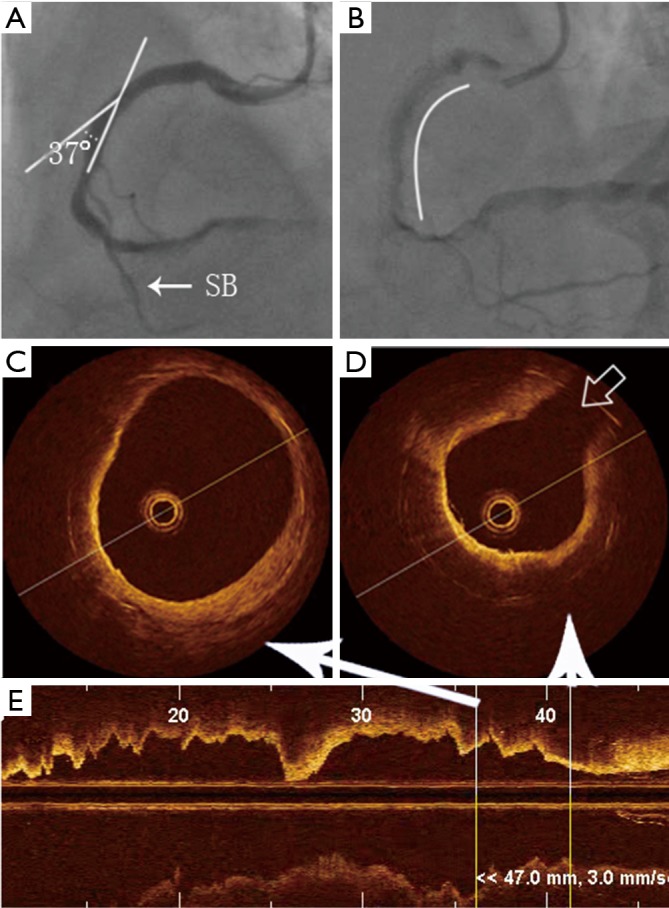
Figure 2.

Localization of ISNA in relation to curvatures and bifurcations by OCT. (A) Angiography showing the vascular angle to be 37°; (B) the stent is in the middle of the RCA, the arc represents the stent length. The distance from the side-branch vessel to the stent edge is 3 mm (SB, side branch); (C,D) example of neoatherosclerosis by OCT, which is identified as a diffusely bordered, signal-poor region with overlying signal-rich homogenous bands. Gray arrow shows the location of the side branch; (E) OCT image shows the longitudinal axis of the cross-section of the blood vessel. ISNA, in-stent neoatherosclerosis; OCT, optical coherence tomography.
