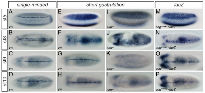
Fig. 1. The sog primary enhancer directs expression in the ventral midline of the late embryo. Approximately 2-10 hours (h) after egg deposition (AED), embryos were collected, dechorionated, and fixed. Whole-mount in situ hybridization was performed with fixed embryos and digoxigenin (DIG)-UTP labeled antisense RNA probes complementary to sim, sog, and lacZ. Each probe used in the individual in situ hybridization is shown on the top of each column. Expression patterns of sim (A-D) were visualized in wild-type (yw) Drosophila embryos. An antisense sog RNA probe was used to target endogenous sog transcripts in both wild-type (yw) (E-H) and sim mutant (sim−/−) (I-L) embryos. The sim mutant embryo was homozygous for the sim2/H9 allele. (M-P) Expression of a lacZ fusion gene directed by a ∼0.4-kb sog primary enhancer in a transgenic embryo recapitulated the endogenous pattern of sog expression in the neurogenic ectoderm and the ventral midline (compare with E-H). ‘st’ indicates the developmental stage of Drosophila embryogenesis. Developmental stages were defined according to previously established criteria (23).

