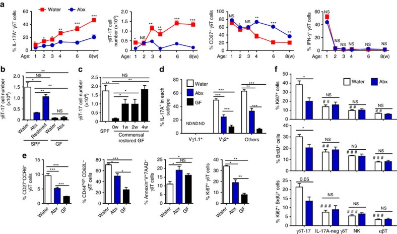
Figure 2. The microbiota maintain the pool of hepatic γδT-17 cells in steady state.
(a) FACS analysis of IL-17A, CD27 and IFN-γ expression by hepatic γδT cells at the indicated B6 mouse ages over time. The mice began treatment with water alone (Water) or containing antibiotics (Abx) in utero through the pregnant mother (n=7/time point). (b) Five-week-old adult SPF B6 mice and GF mice were fed water alone (Water) or containing antibiotics (Abx) for 4 weeks. Abx-pretreated SPF mice were co-housed with normal mice for an additional 4 weeks to reconstitute their gut bacteria (Restored), and the hepatic γδT-17 cell numbers were evaluated by FACS (n=4 for SPF mice, n=5 for GF mice). (c) Ten-week-old GF mice were co-housed with SPF mice to restore their commensal microbes, and the hepatic γδT-cell number was detected at the indicated time points post co-housing (n=9, 4, 5, 5, 10; from left to right). The frequency of IL-17A expression by each hepatic γδT-cell subtype (n=10, 7, 5; from top to bottom) (d) and the frequency of hepatic γδT cells with the phenotype CD44highCD62L−, CD27−CCR6+, Annexin-V+7AAD− and Ki67+ (n=5 per group) (e) from Water-treated mice, Abx-treated mice and GF mice were analysed by FACS. (f) Control and Abx-treated mice were i.p. injected with 1 mg BrdU three times at 2-day intervals, and BrdU incorporation and Ki67 expression by hepatic γδT-17 (CD3+γδTCR+IL-17+), IL-17A negative γδT (CD3+γδTCR+IL-17−), NK (CD3–NK1.1+) and αβT (CD3+TCRβ+) cells were evaluated by FACS. Differences between γδT-17 cells and other cells from control mice (indicated as ‘#’) and differences between Water- and Abx-treated mice of each subtype of cells (indicated as ‘*’) are shown (n=6, 4; Water, Abx). The data are representative of more than three independent experiments. The mean±s.e.m. is shown (*P<0.05; **,##P<0.01; ***,###P<0.001 one-way ANOVA with post hoc test.).

