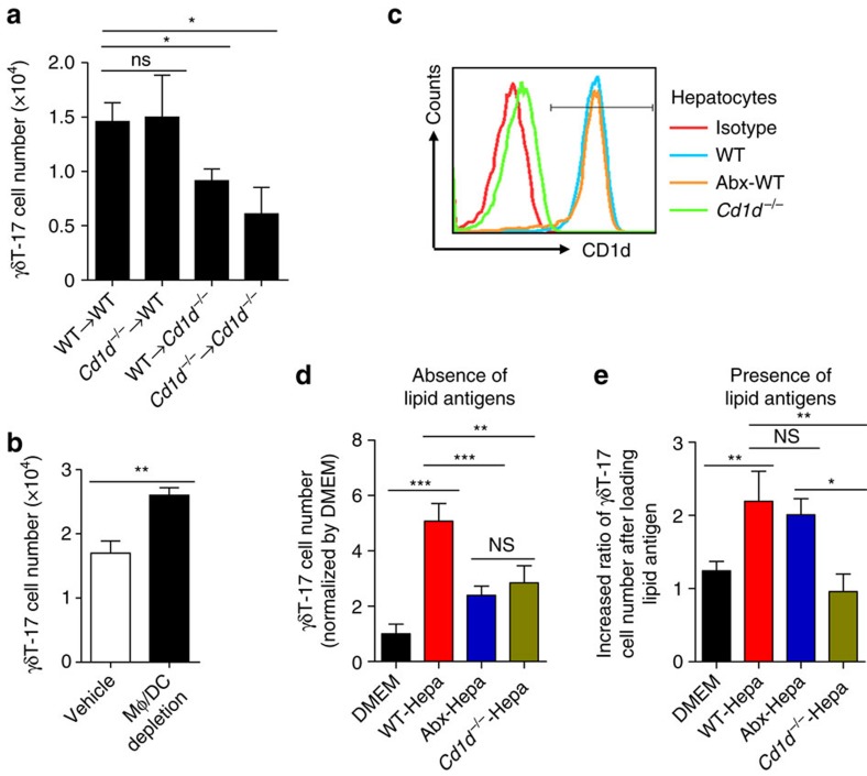
Figure 6. Hepatocytes loaded with lipid antigen promote hepatic γδT-17 cells in vitro.
(a) Quantification of hepatic γδT-17 cells from chimeric mice reconstructed with fetal liver MNCs (n=4 per group). (b) The numbers of hepatic γδT-17 cells from vehicle- or clodronate liposome-treated WT mice (n=4, 5). (c) CD1d expression on the indicated hepatocytes. (d) Purified hepatic γδT cells from WT mice were co-cultured with the indicated hepatocytes for 3 days, and the γδT-17 cell number (normalized by the DMEM group) was analysed by FACS (n=6 wells per group). (e) Purified γδT cells were co-cultured with lipid antigen-loaded hepatocytes for 3 days. The increased ratios of γδT-17 cell numbers compared with those without added lipid antigens are shown (n=6 wells per group). The data are representative of three independent experiments and shown by the mean±s.e.m. (*P<0.05; **P<0.01; ***P<0.001 unpaired Student's t-test (b), one-way ANOVA with post hoc test (a,d,e).

