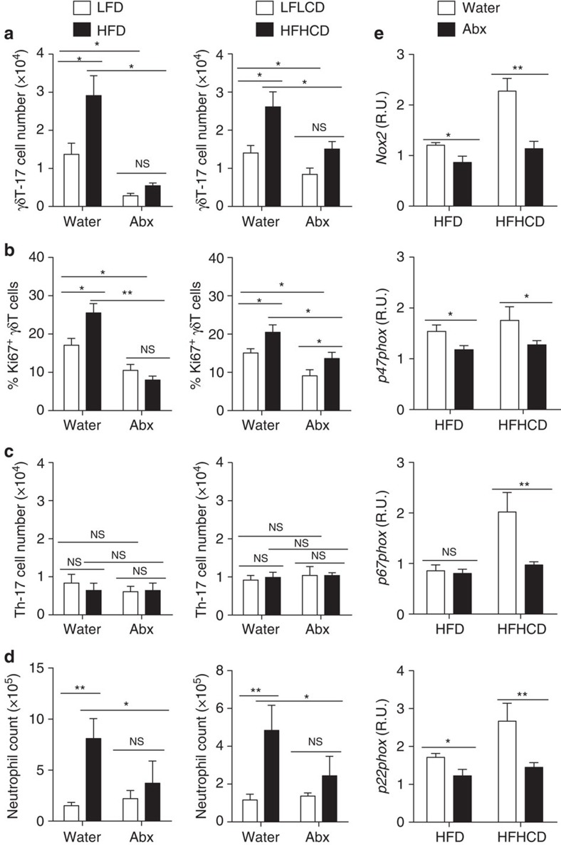
Figure 8. The microbiota promote the expansion of liver-resident γδT-17 cells during HFD/HFHCD-induced NAFLD.
Normal water-fed mice and Abx-treated B6 mice were placed on an HFD (LFD as control) and an HFHCD (LFLCD as control) for 24 weeks. (a) The hepatic γδT-17 cell number, (b) Ki67 expression of hepatic γδT cells, (c) hepatic Th-17 cell number and (d) neutrophil number in the liver were detected by FACS. (e) Water- and Abx-treated B6 mice were placed on an HFD or an HFHCD for 12 weeks. The hepatic mRNA expression levels of Nox2, p47phox, p67phox and p22phox were evaluated. The data are representative of three independent experiments with five mice per group and shown by the mean±s.e.m. (*P<0.05; **P<0.01; ***P<0.001 one-way ANOVA with post hoc test.).

