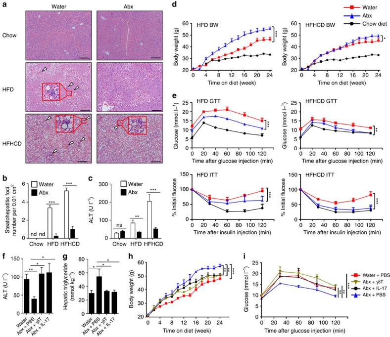
Figure 9. The microbiota accelerate HFD/HFHCD-induced NAFLD through hepatic γδT-17 cells.
(a–e) Mice were treated as described in Fig. 8. (a) Representative liver histology (H&E staining, scale bar, 200 μm); arrowheads indicate the steatohepatitis foci of inflammation with clusters of inflammatory cells, and the red rectangle indicates a 4 × zoom of the gated region. (b) Numbers of steatohepatitis foci counted in a (n=5 per group). (c) Serum ALT level, (d) body weight curve and (e) glucose tolerance test (GTT) and insulin tolerance test (ITT) curves are shown (n=5 per group). (f–i) HFD-fed, Abx-treated mice were either injected with IL-17A (500 ng, once per week) or transferred with hepatic γδT cells (2 × 10⁴, once per 2 weeks) from the 4th week to the 24th week of HFD treatment, (f) The serum ALT level, (g) hepatic triglyceride level, (h) body weight curve and (i) GTT curve are shown (n=5 per group). The data are representative of three independent experiments and shown by the mean±s.e.m. (*P<0.05; **P<0.01; ***P<0.001 one-way ANOVA with post hoc test (b,c,f,g), two-way ANOVA test (d,e,h,i).

