Figure 5.
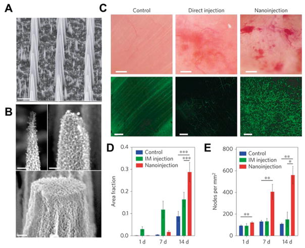
Nanoneedle for the delivery of VEGF-165 to upregulate blood vessel formation in muscle. (A) Scanning electron microscope (SEM) micrographs showing the morphology of porous silicon nanoneedle arrays with pitches of 2 μm. Scale bars, 2 μm. (B) High-resolution SEM micrographs of nanoneedle tips showing the nanoneedles’ porous structure and the tunability of tip diameter from less than 100 nm to over 400 nm. Scale bars, 200 nm. (C) Intravital bright-field (top) and confocal (bottom) microscopy images of the vasculature of untreated (left) and hVEGF-165-treated muscles with either direct injection (center) or nanoinjection (right). The fluorescence signal originates from systemically injected FITC–dextran. Scale bars, bright-field 100 μm; confocal 50 μm. (D, E) Quantification of the fraction of fluorescent signal (dextran) (D) and the number of nodes in the vasculature per mm2 (E) within each field of view acquired for untreated control, intramuscular injection (IM) and nanoinjection. *p = 0.05, **p < 0.01, ***p < 0.001. Error bars represent the SD of the averages of five areas taken from three animals. Reprinted with permission of figure and caption text from ref 48. Copyright 2015 Macmillan Publishers Ltd.

