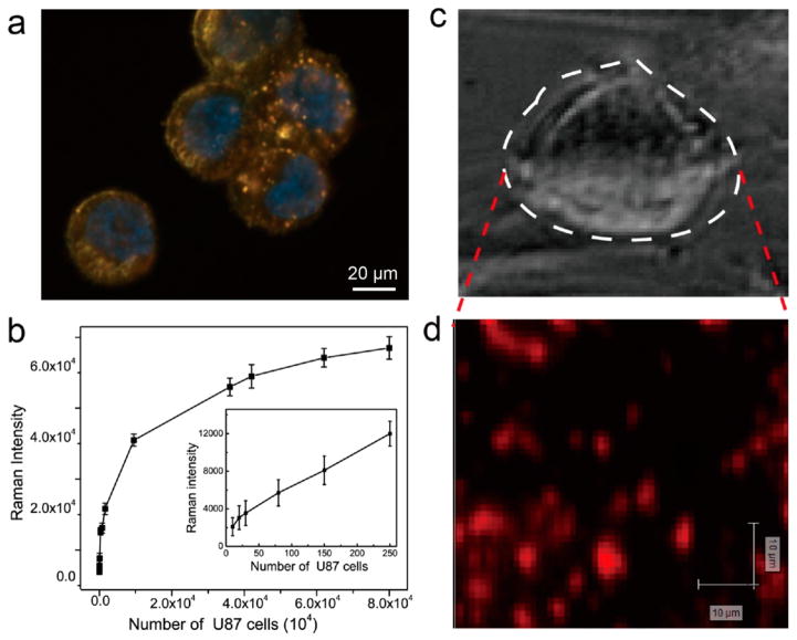
Figure 5.
(a) Dark-field images of U87MG cells treated with PEGylated CNTR@AuNP. The cell nuclei were counterstained with Hoechst 3342, exhibiting blue fluorescence. (b) Raman intensity at 1588 cm−1 as a function of cell density (cells/mL). (c) Bright-field image and (d) Raman mapping of a single U87MG cancer cell incubated with PEGylated CNTR@AuNP.

