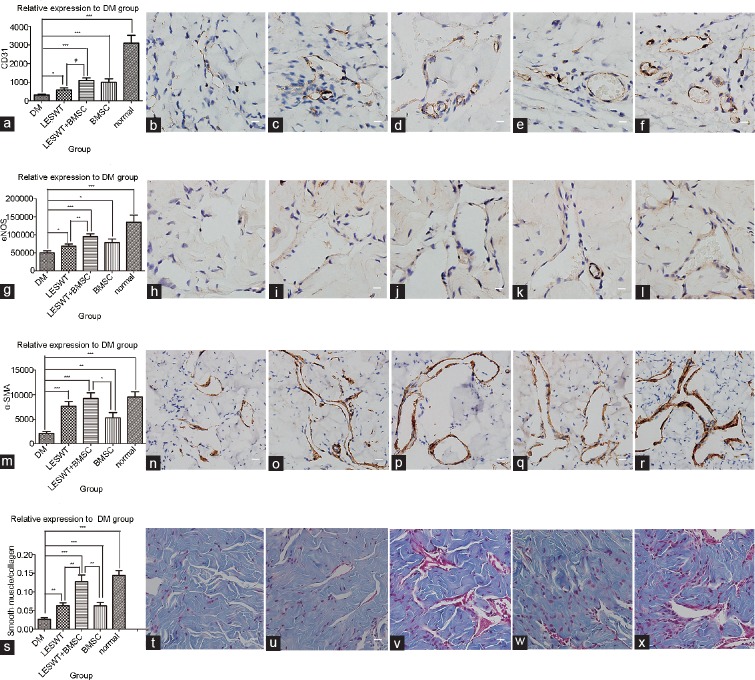
Figure 6.
Immunohistochemistry of the cavernous tissue. Brown dye indicated the target factor-positive expression area. CD31, eNOS, or α-SMA-positive stained cells identified in the cavernous body appeared mostly on the inner wall surface of the vasculature or inside the vessel walls. The average stained area was expressed relative to the DM group. Compared with the normal group, a significant decrease in the positively stained area of CD31, eNOS, α-SMA or smooth muscle/collagen ratio was observed in the DM group, suggesting serious cell damage and fibrosis in penile tissue. After diabetic rats were treated by LESWT or BMSC transplantation, CD31-, eNOS-, and α-SMA-positive stained areas and smooth muscle/collagen ratio increased significantly. The combination of LESWT and BMSC transplantation increased CD31 and eNOS expression more effectively than LESWT, increased α-SMA expression more effectively than BMSC transplantation, and increased the smooth muscle/collagen ratio more effectively than LESWT and BMSC transplantation performed alone. (a–f) CD31 expression in the cavernous tissue. (g–l) eNOS expression in the cavernous tissue. (m–r) α-SMA expression in the cavernous tissue. (s–x) Smooth muscle/collagen ratio in the cavernous tissue. (a, g, m and s) Cell markers relative expression to DM group. (b, h, n and t) DM group. (c, i, o and u) LESWT group. (d, j, p and v) LESWT + BMSC group. (e, k, q and w) BMSC group. (f, l, r and x) Normal group. *P < 0.05; **P < 0.01; ***P < 0.001. Scale bar: 25 µm. eNOS: endothelial nitric oxide synthase; α-SMA: α-smooth muscle actin; DM: diabetes mellitus; LESWT: low-energy shock-wave therapy; BMSC: bone marrow mesenchymal stem cell.

