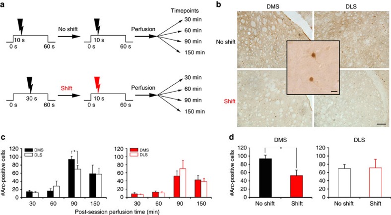
Figure 5. Arc expression is reduced in the DMS when the CS–US interval is shifted.
(a) Schematic explaining experimental design in test (shift) and control (no shift) groups. The ‘shift' group underwent an aversive conditioning protocol with a CS–US interval of 30 s until the day of shift where such interval was shortened to 10 s. The control group was trained with a fixed CS–US interval of 10 s all along. With this design, the session animals underwent before perfusion was strictly the same for both groups. The only difference resides in the fact that the 10 s interval is a new duration for one group, and not for the other. Animals were perfused at different times after the last behavioural session (no-shift, n=6 per time point; shift, n=5 per time point). (b) Photomicrographs of transverse Arc-stained sections from representative cases illustrating expression in the DMS and DLS 90 min after the shifted versus non-shifted session. Scale bar, 200 μm. The inset shows a high-resolution image of DMS neurons immunostained for Arc. Scale bar, 10 μm. (c) Quantification of the number of striatal Arc-positive cells (mean+s.e.m.) for non-shifted (left panel) or shifted (right panel) animals perfused at different time points after the behavioural session revealed that Arc expression 90 min post-training was higher in the DMS than in the DLS for the non-shifted animals (ANOVA, *P=0.03). (d) Quantification of the number of striatal Arc-positive cells (mean+s.e.m.) for DMS and DLS in non-shifted and shifted animals perfused at 90 min after the behavioural session shows a reduction in Arc expression in the DMS of shifted rats. ANOVA, *P=0.02.

