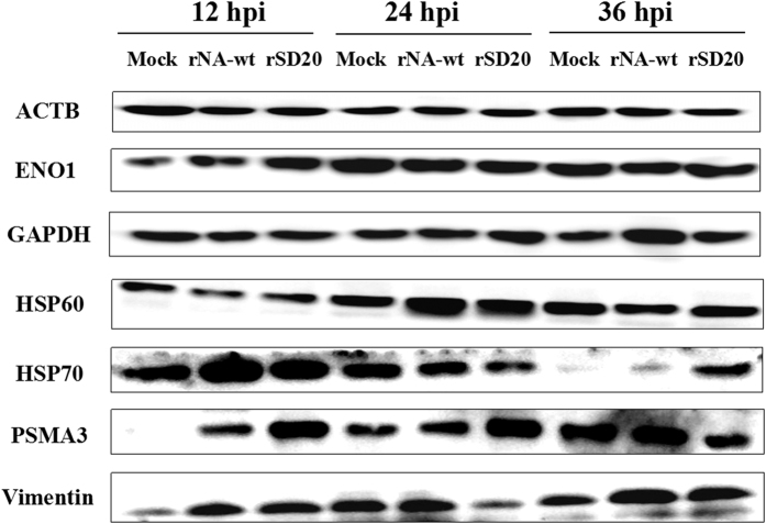
Figure 4. Western blot analysis of selected proteins.
The proteins were detected from individual samples of CEF cells infected by rNA-wt, rSD20 or PBS that were collected at 12, 24 and 36 hpi. The inoculations are indicated at the top and the names of the six DE proteins, together with β-actin, are shown on the left. Full-length blots/gels are presented in Supplementary Figs S4~S10 respectively.

